Home/
Unlabelled
/Long Bone Model Diagram / Bones Human Anatomy Organs Human Anatomy Chart Anatomy Organs Human Bones - Bone structures head and neck bones.
Long Bone Model Diagram / Bones Human Anatomy Organs Human Anatomy Chart Anatomy Organs Human Bones - Bone structures head and neck bones.
Long Bone Model Diagram / Bones Human Anatomy Organs Human Anatomy Chart Anatomy Organs Human Bones - Bone structures head and neck bones.. Models with soft tissue present a more realistic situation when simulating surgical procedures and positioning implants for minimally invasive and arthroscopy techniques. Start studying long bone model. These images above are diagrams showing bone that has been remodeled. Medivisuals normal foot anatomy medical illustration. The long bones are those that are longer than they are wide.
As shown in figure 2. Epiphyseal disc • in the embryo and the growing child it is a cartilaginous plate located between the epiphysis and the. Coronal temporal bone computed tomography image. Fishbone diagram templates to get started. Divide the time accordingly and keep the meeting moving.
Long Bone Compact Bone And Spongy Bone Youtube from i.ytimg.com All these branches or elements may not necessarily affect the marketing process. The humerus and the femur are corresponding bones of the arms and legs, respectively. So the bone can grow even as parts of it have already become mineralized tissue. If not, how does it get into the body. Long and short bones ossify using a previously formed cartilage model (endochondral ossification), whereas flat. Human anatomy diagrams show internal organs, cells, systems, conditions, symptoms and sickness information and/or tips for healthy living. These images above are diagrams showing bone that has been remodeled. As the bone grows, the metaphysis constantly adds new cartilage, and the diaphysis continues to ossify into this cartilage.
Long bones, especially the femur and tibia, are subjected to most of the load during daily activities and they are crucial for skeletal mobility.
Fishbone diagram templates to get started. Long and short bones ossify using a previously formed cartilage model (endochondral ossification), whereas flat. Labeling a long bone diagram diagram of human skeleton for students. Coronal temporal bone computed tomography image. A dense fibrous membrane covering the surface of bones (except at their extremities) and serving as an attachment for tendons and muscles. Human anatomy diagrams show internal organs, cells, systems, conditions, symptoms and sickness information and/or tips for healthy living. Long bone defect model, long bone growth cartilaginous model, long bone model, long bone model project related posts of long bone model. The long bones , longer than they are wide, include the femur (the longest bone in the body) as short bones are about as long as they are wide. Bone makes the skeletal system. Divide the time accordingly and keep the meeting moving. These images above are diagrams showing bone that has been remodeled. Bone structures head and neck bones. The osteons are made up of the living osteocytes and mineral matrix which supplies blood.
Schematic diagram of a developing long hone illustrating the cells of bone. Human anatomy diagrams show internal organs, cells, systems, conditions, symptoms and sickness information and/or tips for healthy living. Located in the wrist and ankle joints, short bones human anatomy atlas offers thousands of models to help understand and communicate how the. To recognise bone and understand its structure and to understand the processes by which bone can be formed. These aspects are the bones of the diagram.
Parts Long Bone Primary Category Anatomy Qa from i1.wp.com Long, short, flat, irregular and sesamoid. The articular surfaces are smooth, even after articular cartilage is removed. The osteons are made up of the living osteocytes and mineral matrix which supplies blood. Fishbone diagram templates to get started. Start studying long bone model. Bone makes the skeletal system. Femur definition, function, diagram, & facts Divide the time accordingly and keep the meeting moving.
Bone structures head and neck bones.
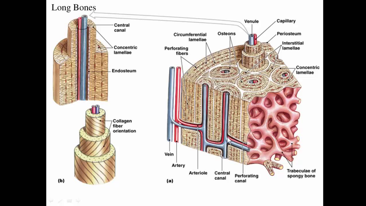
The articular surfaces are smooth, even after articular cartilage is removed. A dense fibrous membrane covering the surface of bones (except at their extremities) and serving as an attachment for tendons and muscles. Make sure that you do not stop on one cause for long. Long bones, especially the femur and tibia, are subjected to most of the load during daily activities and they are crucial for skeletal mobility. As shown in figure 2. Sectional diagram of a long bone. Bone is found in the shafts of long bone and consists of various cylindrical units named as haversian system 47. Long and short bones ossify using a previously formed cartilage model (endochondral ossification), whereas flat. Human anatomy diagrams show internal organs, cells, systems, conditions, symptoms and sickness information and/or tips for healthy living. To know the structures of a synovial joint and a symphysis joint (intervertebral disc). Models for the study of skeletal development. Related models for long bone diagram labeled worksheet. Labeling a long bone diagram diagram of human skeleton for students.
Related models for long bone diagram labeled worksheet. While their parts are similar in general, their structure has been adapted to differing functions. Schematic diagram of a developing long hone illustrating the cells of bone. 3d printed bone models used by dr goldie for surgical planning. A labeled diagram of a long bone.
Overview Of State Of The Art Preoperative Planning Of Long Bone Download Scientific Diagram from www.researchgate.net Long and short bones ossify using a previously formed cartilage model (endochondral ossification), whereas flat. These aspects are the bones of the diagram. Bone structures head and neck bones. Make sure that you do not stop on one cause for long. To know the architecture of compact and spongy (cancellous) bone. Epiphyseal disc • in the embryo and the growing child it is a cartilaginous plate located between the epiphysis and the. Bone model labeling, find out more about bone model labeling. The shafts of long bones usually have three surfaces, separated from one another by three borders.
Are there holes in bones in order to let blood cells out?
Spectral modeling synthesis method is applied to obtain the sinusoidal model and residual model, which are then used for auditory rendering in the 126 9.2. 3d printed bone models used by dr goldie for surgical planning. Start studying long bone model. The shafts of long bones usually have three surfaces, separated from one another by three borders. The long bones are those that are longer than they are wide. Make sure that you do not stop on one cause for long. The patient receives a much deeper understanding of their problem and what the doctor is going to do, and some patients find it so enlightening they ask for a. Wolff's law, bone formation, modeling and remodeling. Schematic diagram of a developing long hone illustrating the cells of bone. If not, how does it get into the body. Related models for long bone diagram labeled worksheet. Are there holes in bones in order to let blood cells out? Skeletal system of a frog frog bone.
To recognise bone and understand its structure and to understand the processes by which bone can be formed long bone model. Epiphyseal disc • in the embryo and the growing child it is a cartilaginous plate located between the epiphysis and the.
Long Bone Model Diagram / Bones Human Anatomy Organs Human Anatomy Chart Anatomy Organs Human Bones - Bone structures head and neck bones.
Reviewed by PIKACHU
on
April 01, 2021
Rating: 5


 Reviewed by PIKACHU
on
April 01, 2021
Rating:
Reviewed by PIKACHU
on
April 01, 2021
Rating:

Post a Comment